Identifying Peripheral Artery Disease
Why is it critical to identify peripheral artery disease (PAD) in patients with foot ulcers?
Among patients foot ulcer or foot gangrene, 1-year rates of leg amputation are 50-60% (see review) if peripheral artery disease (PAD) is untreated or unrecognized. In contrast, 1-year leg amputation rates following revascularization are 90%.
Thus, the identification and treatment of PAD in patients with foot ulcers may be the single most influential factor in amputation prevention. Revascularization is cost-effective and may even provide cost-savings in frail, elderly patients.
An evidence-based approach to testing
There currently is no non-invasive test that has perfect diagnostic accuracy for PAD. Because of the negative consequences on limb outcomes that occur when PAD is missed, the ideal strategy toward testing should be:
- An initial screening test with 100% sensitivity.
- A confirmatory test with 100% specificity.
This is exactly the same strategy with other high-stakes testing done in health care (ex. COVID) and in non-health care situations (ex. pre-boarding screening at airports).
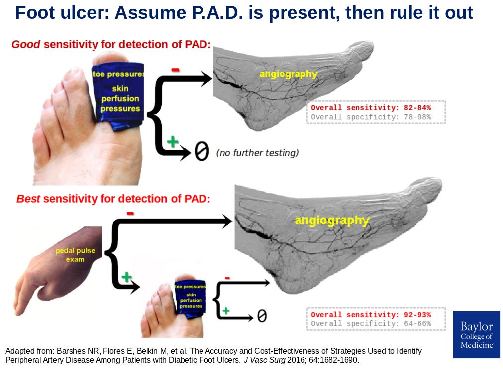
These two formal decision analysis identified these as the two sensitive strategies to identify PAD in patients with foot ulcers.
The best diagnostic strategy
Feel for pedal pulses.
- If at least one pedal pulse is NORMAL/2+ PALPABLE: confirm with non-invasive testing to corroborate adequate arterial perfusion. Angiography if toe-brachial index >0.7 (see below).
- If pedal pulses are WEAK or ABSENT: angiography without the need for non-invasive testing.
This strategy has a 92% sensitivity (i.e. misses the diagnosis of PAD in only about one out of every 13 patients with PAD.
In addition, consider angiography without need for non-invasive testing if any of the following specific situations are present:
| finding | |—| | foot ulcer in area supplied by single angiosome: heel, dorsal foot | | anticipated Lisfranc amputation, rotational flap, or free flap reconstruction | | ulcer size >3cm | | infection requiring >3cm incision for drainage | | ulcers in multiple locations/angiosomes | —
An acceptable diagnostic alternative
Non-invasive testing for all patients.
-
Angiogram to investigate abnormal results = 83% sensitivity (i.e. misses the diagnosis of PAD in about one out of every six patients with PAD.
Findings from the decision analysis suggest that reserving non-invasive testing for use only in further evaluation of patients with weak or absent pedal pulses has a very LOW sensitivity rate in finding PAD. This may be at least partly explained by the poor diagnostic accuracy of palpating for pedal pulses in diabetic feet.
In summary, all patients with foot ulcers should get objective testing, either in the form of an angiogram or non-invasive testing. Pedal pulse exam alone is INSUFFICIENT to reliably rule out PAD.
Support for the toe-brachial index
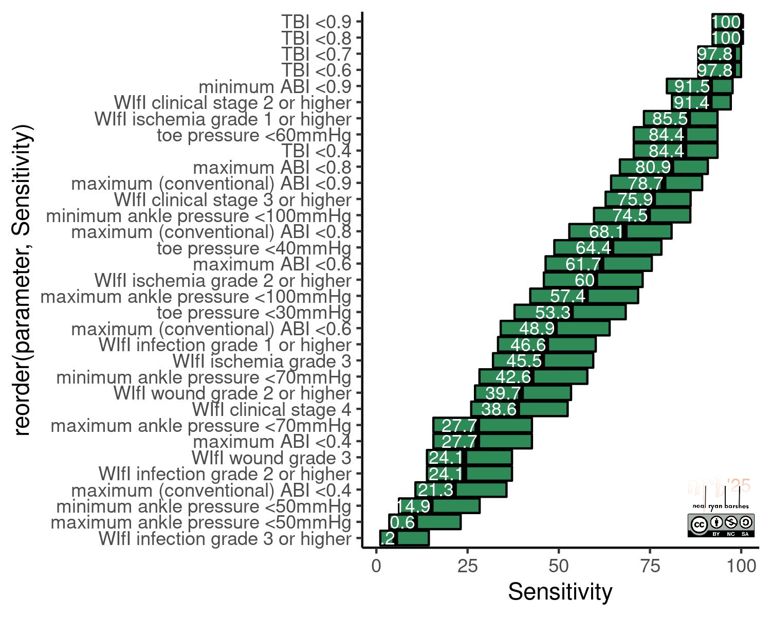
Since performing that formal decision analysis, we have corroborated findings in a study that compared angiographic with non-invasive testing results on 100 consecutive patients with non-healing ulcers and suspected PAD. PAD severity as seen on digital subtraction angiography was quantified independently by two board-certified vascular surgeons using the Society for Vascular Surgery Global Limb Anatomic Staging System (GLASS) classification system. Non-invasive pressure testing was done by registered vascular technicians in an ICAVL-approved vascular lab. Medial artery calcification was quantified on foot radiographs.
The most important finding of this analysis:
Toe-brachial index of 0.7 was the single most sensitive testing threshold to identify GLASS 2 or 3 (moderate/severe) PAD. This parameter outperformed absolute pressure thresholds and WIfI ischemia grading. This parameter was also affected far less by medial artery calcification of foot arteries.
Additional resources
For some further explanation, consider these brief videos on bedside evaluation and non-invasive lower extremity arterial testing for PAD. For further detail still, read this chapter on non-invasive testing for peripheral artery disease.